

TL;DR:
- Dental barrier function is a multi-layered defense system formed by the gums and oral mucosa that prevents bacterial invasion and maintains tissue health. When this system is compromised by inflammation or damage, it can lead to periodontal disease and systemic health issues. Proper oral hygiene, professional care, and supportive products strengthen and preserve this dynamic biological barrier.
Dental barrier function is defined as the protective and immunological seal formed by the gums and oral mucosa that prevents bacterial invasion, supports tissue integrity, and reduces the risk of periodontal disease. This seal is not a passive wall. It is a multilevel defense system that includes physical, chemical, microbial, and cellular components, each remodeling dynamically in response to infection, inflammation, and environmental stress. Understanding dental barrier function is the first step toward recognizing why consistent oral hygiene does far more than keep teeth clean. It preserves a biological system that also protects systemic health, including cardiovascular and metabolic function.
What is dental barrier function and how is it structured?
The gingival barrier is defined as the protective seal of soft tissue surrounding each tooth, acting as the primary physical and immune defense against bacterial entry. It serves two simultaneous roles: blocking pathogens mechanically and coordinating an active immune response. These two functions are not separate. They operate as an integrated system.
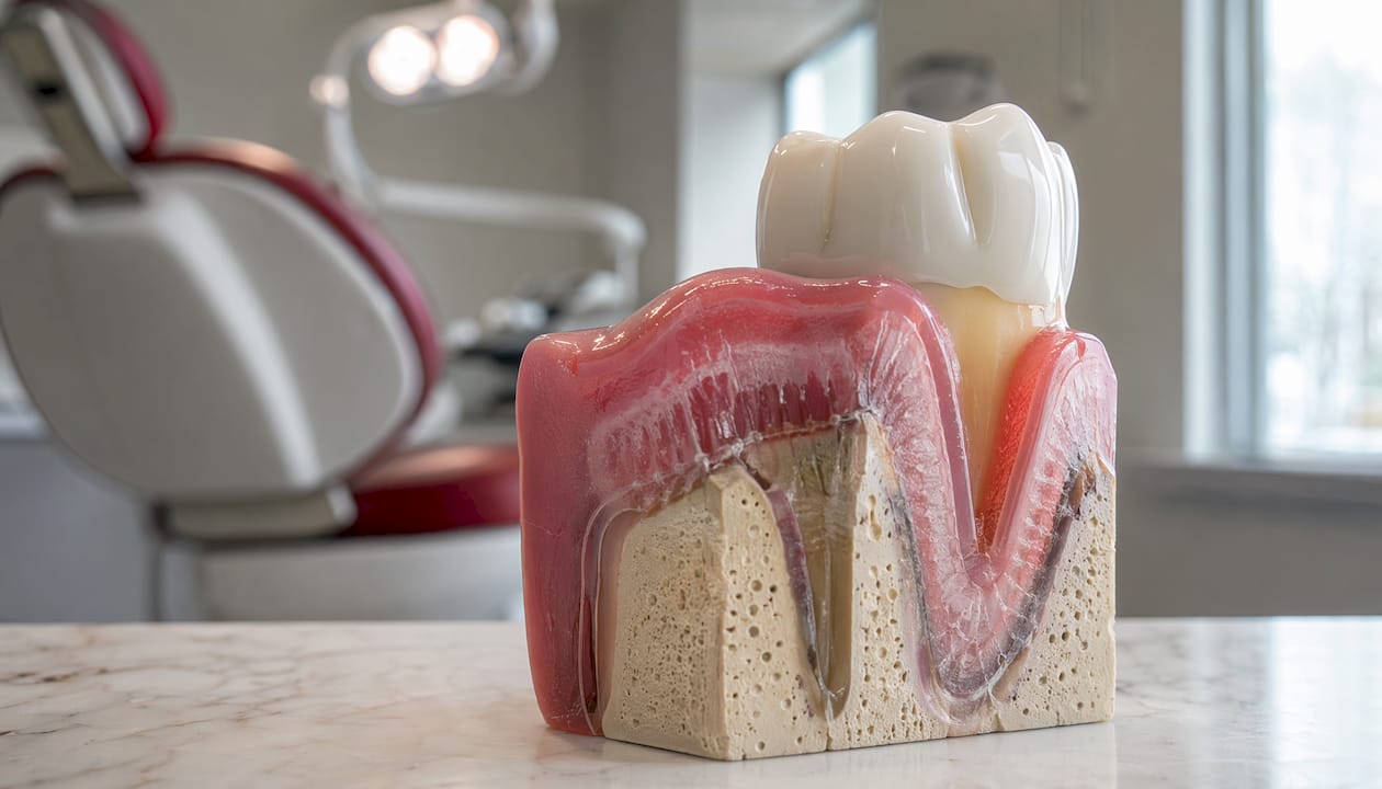
The oral mucosal barrier includes the gingival tissue, the junctional epithelium, and the underlying connective tissue. Together, these layers form a continuous seal between the external oral environment and the deeper periodontal structures. When this seal is intact, bacteria in dental biofilm cannot penetrate the tissue and trigger destructive inflammation.
Saliva reinforces this seal from the outside. It contains antimicrobial peptides and proteins, including lysozyme, lactoferrin, and defensins, that neutralize pathogens before they reach the epithelial surface. Mucins in saliva form a hydrated gel that physically traps microbes. This chemical and mechanical layer is the first line of defense before bacteria even contact gum tissue.
What anatomical components form the dental barrier?
The dental barrier draws its strength from several distinct anatomical structures, each contributing a specific protective role.
- Gingival epithelium: The outermost cellular layer of the gums. It forms a physical wall against bacterial penetration and acts as an immune sentinel, detecting pathogens and signaling immune responses.
- Junctional epithelium: The specialized tissue that attaches the gum to the tooth surface. It is more permeable than surface epithelium, which allows immune cells to migrate through it, but this permeability also makes it a vulnerable entry point when compromised.
- Supracrestal gingival tissue (SGT): This tissue forms a critical immunobiological barrier at the base of the gingival sulcus, providing structural support and immune surveillance directly above the alveolar bone.
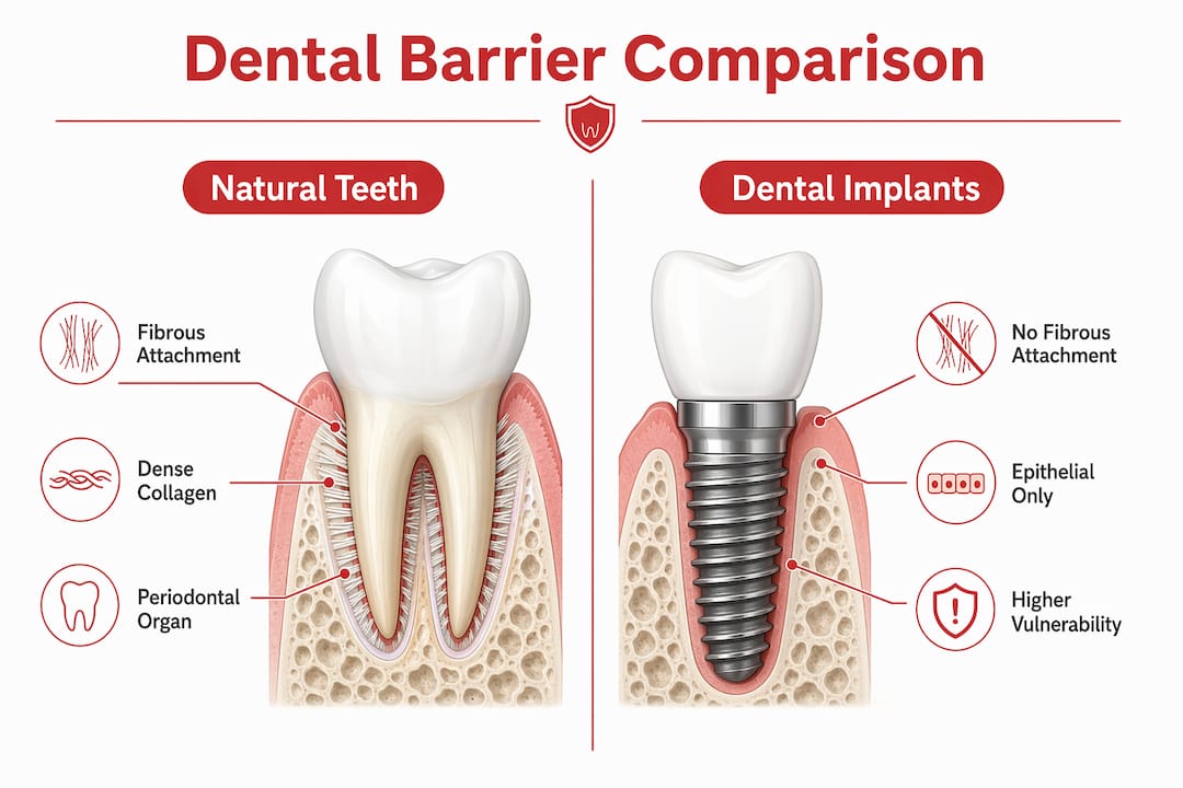
- Periodontal organ (PO): The dense collagen fibrous barrier of the periodontal organ provides mechanical strength and anchors the tooth within the socket. This structure is unique to natural teeth.
- Immune cells: Neutrophils, macrophages, and dendritic cells are embedded throughout gingival tissue. They patrol for bacterial antigens and coordinate inflammatory responses when the barrier is breached.
Dental implants lack the periodontal organ entirely. Peri-implant tissues rely solely on epithelial attachment, which provides significantly weaker protection against bacterial invasion. This structural difference explains why implants require more rigorous maintenance protocols than natural teeth.
Pro Tip: If you have dental implants, ask your dentist about peri-implant probing depth at every checkup. Early detection of epithelial detachment is the most effective way to prevent peri-implantitis.
How does the dental barrier protect against bacterial invasion?
The protective mechanisms of the dental barrier operate across four distinct levels, each reinforcing the others.
- Physical exclusion: Intact epithelial cells form tight junctions that physically block bacterial passage. The junctional epithelium, while more permeable, still restricts the movement of large microbial aggregates and their toxins.
- Chemical neutralization: Salivary antimicrobial peptides, including defensins and lysozyme, degrade bacterial cell walls and disrupt biofilm formation before pathogens contact gingival tissue. This chemical layer continuously renews itself through salivary flow.
- Immune surveillance: Gingival epithelial cells actively respond to inflammation through proliferation, migration, and immune activation. They are not passive structural cells. They function as immune sentinels that detect bacterial antigens and recruit neutrophils and macrophages to the site of threat.
- Microbial competition: Commensal oral bacteria occupy surface niches and produce metabolites that inhibit pathogen colonization. A balanced oral microbiome consolidates barrier integrity by preventing dysbiotic species from gaining a foothold.
Periodontal homeostasis is the state in which these four mechanisms operate in balance. Bacterial load remains controlled, immune activation stays proportionate, and tissue remodeling proceeds without net destruction. When any one mechanism weakens, the others compensate up to a threshold. Beyond that threshold, disease begins.
Pro Tip: Gingival epithelial cells renew approximately every 5–7 days. Consistent brushing and flossing supports this renewal cycle by removing biofilm before it matures into a dysbiotic community that overwhelms the barrier.
What happens when the dental barrier is compromised?
Barrier disruption follows a predictable sequence. Infection, chronic inflammation, trauma, or systemic conditions increase epithelial permeability. Bacteria and their toxins penetrate deeper tissue layers. The immune response escalates, and the resulting inflammation begins to damage the very structures it is meant to protect.
Elevated inflammatory cytokines, including IL-1β, IL-6, and TNF-α, impair barrier integrity by modifying tight junction proteins and increasing tissue permeability. This creates a cycle where inflammation worsens the barrier defect, which allows more bacterial penetration, which drives more inflammation. Matrix metalloproteinase (MMP) activity accelerates this process by degrading the collagen fibrous barrier, leading to alveolar bone loss.
The systemic consequences are significant. Barrier disruption is associated with cardiovascular and metabolic diseases, because bacteria and inflammatory mediators that breach the oral barrier can enter systemic circulation. Tooth loss compounds the structural damage. Alveolar ridge width can decrease by up to 50% within 12 months of tooth extraction, reflecting how quickly the supporting architecture collapses once the barrier is gone.
The table below summarizes key differences in barrier vulnerability between natural teeth and dental implants.
| Feature | Natural teeth | Dental implants |
|---|---|---|
| Fibrous attachment | Dense collagen periodontal organ | Absent; epithelial attachment only |
| Barrier strength | High mechanical and immune defense | Reduced; more susceptible to infection |
| Response to inflammation | Active immune modulation via SGT | Limited; relies on peri-implant epithelium |
| Risk of bone loss | Lower with intact barrier | Higher; peri-implantitis progresses faster |
| Maintenance requirement | Standard hygiene protocols | More frequent professional monitoring |
Understanding this vulnerability difference matters for anyone with implants. The gum immune support required around implants is categorically different from what natural teeth need.
What clinical approaches maintain or restore dental barrier function?
Maintaining the dental barrier requires both professional care and consistent daily practice. The following approaches address barrier health at multiple levels.
- Mechanical plaque control: Brushing twice daily and flossing once daily removes biofilm before it matures. Mature biofilm, also called dental biofilm, shifts toward dysbiotic species that actively degrade the epithelial barrier.
- Professional scaling and root planing: Removes calcified deposits that harbor pathogenic bacteria below the gumline. This procedure directly reduces the bacterial load driving chronic barrier inflammation.
- MMP inhibitor therapy: MMP inhibitors show clinical promise in preserving barrier integrity by blocking the collagen-degrading enzymes that accelerate periodontal tissue breakdown. Sub-antimicrobial dose doxycycline is the most studied agent in this category.
- Barrier membrane use in bone regeneration: In cases of significant bone loss, barrier membranes physically separate soft tissue from bone defects, allowing guided bone regeneration. Clinical data shows success rates of 90.6–100% for specific applications such as radicular crack treatment.
- Nutritional support: Vitamin C deficiency directly impairs collagen synthesis in the periodontal organ. Adequate zinc and vitamin D intake supports epithelial cell renewal and immune function within gingival tissue.
- Smoking cessation: Smoking suppresses neutrophil function and reduces salivary antimicrobial peptide output, impairing two of the four protective mechanisms simultaneously.
Natural oral care products formulated to support salivary chemistry and gingival health can complement these clinical approaches. Products that avoid disrupting the oral microbiome are particularly relevant, given that commensal bacteria are an active component of barrier defense.
Key Takeaways
The dental barrier is a dynamic, multi-component system that requires consistent mechanical hygiene, immune support, and microbial balance to function effectively against bacterial invasion and systemic disease.
| Point | Details |
|---|---|
| Barrier definition | The gingival and mucosal seal that physically and immunologically blocks bacterial entry into periodontal tissue. |
| Cellular role | Gingival epithelial cells are active immune sentinels, not passive structural cells, and coordinate inflammatory responses. |
| Implant vulnerability | Dental implants lack the collagen fibrous periodontal organ, making them significantly more susceptible to infection. |
| Systemic risk | Barrier disruption links to cardiovascular and metabolic disease through systemic bacterial and cytokine translocation. |
| Maintenance priority | Daily plaque removal, professional scaling, and microbiome-supportive oral care are the most effective barrier preservation strategies. |
The barrier is more dynamic than most patients realize
When I trained in dentistry, the gingival barrier was taught primarily as a structural concept. The emphasis was on tissue anatomy, probing depths, and attachment levels. What that framework missed was the immunological intelligence embedded in the tissue itself.
The shift I have seen in clinical research over the past decade is significant. Gingival epithelial cells are now understood to function as immune sentinels, not passive walls. They detect bacterial antigens, modulate cytokine release, and coordinate the recruitment of neutrophils and macrophages. That is not passive barrier function. That is active immune participation.
What this means clinically is that patient education needs to change. Telling patients to “brush and floss” is accurate but incomplete. The goal is to support a living biological system, not just clean a surface. When patients understand that their gums contain immune cells actively defending their teeth, they engage with oral hygiene differently. They understand why disrupting the oral microbiome with harsh antiseptics can backfire, and why consistent, gentle care outperforms aggressive but irregular intervention.
The natural medicine perspective I bring to this work reinforces that conclusion. Products formulated to work with the oral microbiome, rather than against it, align with what the science now shows about how the barrier actually functions. The barrier is not a wall to be scrubbed. It is an ecosystem to be maintained.
— Veronica
How Stop-oralcare supports dental barrier health
Stop-oralcare develops oral care products formulated with hemp and Dead Sea minerals, ingredients selected for their compatibility with the oral microbiome and gingival tissue. The approach reflects the science: supporting barrier function means working with the body’s existing defense mechanisms, not overriding them.
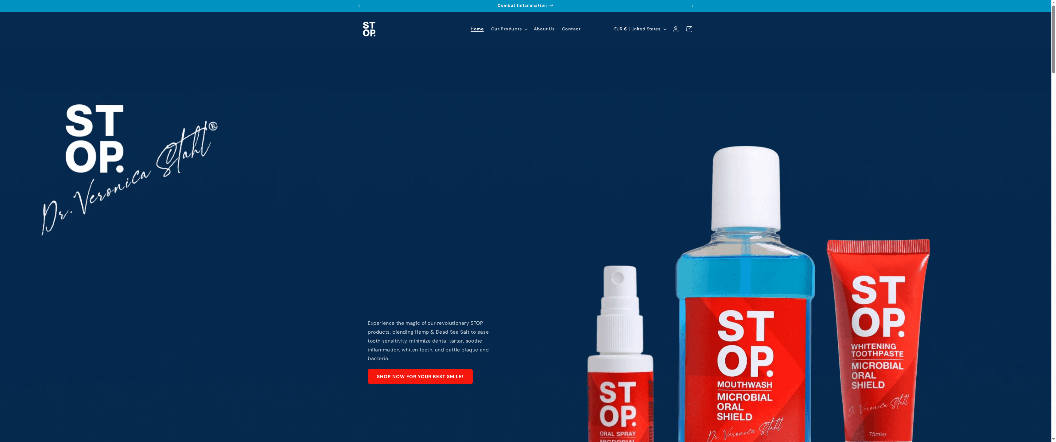
The Stop-oralcare product line includes fluoride-free toothpaste, mouthwash, and oral sprays designed to maintain salivary chemistry and reduce dysbiotic bacterial load without disrupting commensal populations. For readers who want to apply the science in this article to their daily routine, the Stop-oralcare oral care range offers a starting point grounded in both clinical research and natural formulation principles. The educational blog also covers related topics in depth, from microbiome health to periodontal disease care.
FAQ
What is dental barrier function in simple terms?
Dental barrier function is the protective system formed by the gums and oral mucosa that blocks bacteria from entering deeper oral tissues. It combines physical, chemical, immune, and microbial defenses working simultaneously.
How do gingival epithelial cells protect the periodontium?
Gingival epithelial cells detect bacterial antigens and actively recruit immune cells to the site of infection, functioning as immune sentinels rather than passive structural tissue. This active role is central to maintaining periodontal homeostasis.
Why are dental implants more vulnerable than natural teeth?
Dental implants lack the dense collagen fibrous periodontal organ that anchors natural teeth and provides strong barrier protection. Peri-implant tissue relies on epithelial attachment alone, which offers significantly weaker defense against bacterial invasion.
What disrupts dental barrier function?
Chronic inflammation, pathogenic bacterial overgrowth, smoking, nutritional deficiencies, and trauma all compromise barrier integrity. Elevated cytokines such as IL-1β and TNF-α directly impair tight junction proteins, increasing tissue permeability and accelerating disease progression.
Can the dental barrier be restored after damage?
Professional scaling, guided bone regeneration with barrier membranes, MMP inhibitor therapy, and consistent daily hygiene can restore significant barrier function. Early intervention produces the best outcomes, as advanced collagen and bone loss are difficult to fully reverse.